|
|
C-Flap |
|
|---|

|
|
|
It is true that Millard's early diagrams showed the tip of C-flap being placed across the nostril sill and sutured to the tip of the nasal alar base5. Since 1964, however, he has repeatedly written about the proper use of C-flap to lengthen the columella2, 10, 11, 12. In spite of this, one still sees lips in which C-flap has been placed as originally drawn.
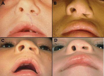
Fig. 60 and Fig. 61 demonstrate the kinds of deformities that result from such behavior: a short columella on the cleft side, an abnormal nostril sill, asymmetric alar bases, an obvious scar along the lower (and sometimes the upper) border of the flap. Lips repaired in this manner have no hope of achieving the true potential of a rotation-advancement repair. |
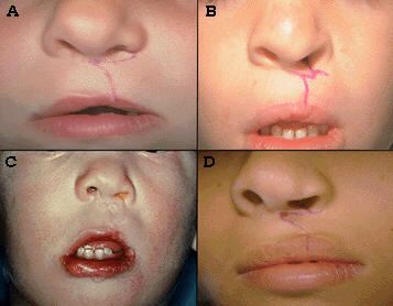
Fig. 60
Fig. 61 |
|
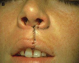
Fig. 62A, an example of a lip being revised, shows pre-operative markings for a R-A repair; not seen is the continuation of C-flap into the nose, continuing up the membranous septum. |
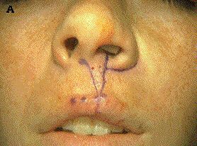
Fig. 62A
|
|
Fig. 62B shows C-flap ready for positioning, the dissection having proceeded up the entire length of the membranous septum, completely freeing medial crus of the lower lateral cartilage in this dissection. C-flap is now part of the ipsilateral hemi-columella anatomic unit. |
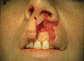
Fig. 62B |
|
Fig. 63A demonstrates the natural position that C-flap assumes when, using a skin hook at the apex of the nostril, one advances the hemi-columella up the membranous septum (where it is sutured in place). |
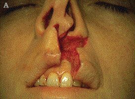
Fig. 63A |
|
Fig. 63B As pointed out by Millard2, 10, 12, this upward columellar advancement leaves a triangular defect on the cleft side of the columella base; it is into this defect that the tip of C-flap is placed. As a result, C-flap provides both increased columellar length and normal flaring of the columellar base. |
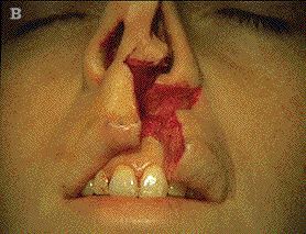
Fig. 63B |
|
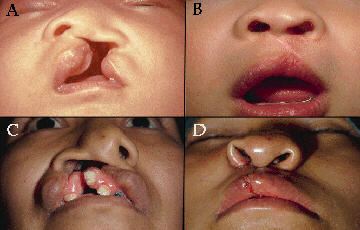
Fig. 64A and B show the pre and post-operative views of this case, demonstrating a more normal post-operative columellar symmetry. |
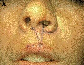
Fig. 64A
Fig. 64B |
|
Fig. 65 and Fig. 66, showing pre-op views on the left and post-op views on the right, illustrate the advantages (i.e., increased columellar length and a normal columellar base) to be achieved when C-flap is correctly used. |
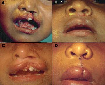
Fig. 65
Fig. 66 |
|
|